(Procyonidae)
Procyonids
Ракунові
Procyonidae is a New World family of the order Carnivora. It includes the raccoons, ringtails, cacomistles, coatis, kinkajous, olingos, and olinguitos. Procyonids inhabit a wide range of environments and are generally omnivorous.
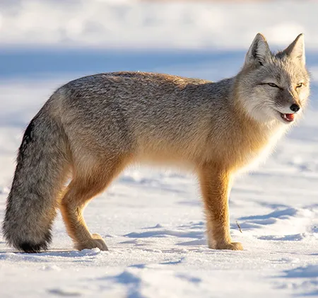
Procyonids are generally small to medium-sized animals, ranging from slightly less than 1 kg to over 20 kg in weight. Some species have slender bodies, while others are stocky. All have medium or long tails. The pelage is gray or brown, sometimes with contrasting markings on the face and light and dark rings around the tail. Most species have relatively short, broad faces; and short but erect ears, which may be rounded or pointed. Forefeet and hindfeet have 5 digits, and procyonids are plantigrade, often walking with a bear-like shuffle. The claws are short and curved. In some species they can be partially retracted. The tail of of species, the kinkajou, is prehensile, and that of coatis is very mobile and is used for balancing during climbing. Males have a well-developed, bilobed baculum.
Procyonid skulls have relatively short rostrums (shorter than canids, longer than felids). They lack alisphenoid canals, but they have well-developed paroccipital processes. Their incisors are unspecialized, and their canines are moderately long and ovate (not round) in cross section. The molars are wide and at least somewhat bunodont. Most species lack secodont carnassials. The dental formula is 3/3, 1/1, 3-4/3-4, 2/2-3 = 36-42.
Most members of Procyonidae are solitary; however, some species form groups. Coati females will form bands of 4 to 24 individuals that forage together, while kinkajous have been found to form social groups of two males and one female. Certain procyonids give birth to one offspring like ringtails, olingos, and kinkajous while raccoons and coatis give birth to litters that range in size from 2 to 6 offspring.
Procyonids are omnivorous. They consume both plant and animal material, including small mammals and birds. Raccoons use their front paws to feel for food items in murky water or leaf litter. Ringtails inspect likely niches and hiding spots in their rocky habitats, hunting for rodents, birds, centipedes, and anything else edible. They are excellent mousers, pouncing and killing with a bite to the back of the rodent’s neck. Coatis dig in the soil and leaf litter using their long claws or their noses to turn up grubs, worms, or other invertebrates. They also turn over large rocks with their front paws to search for invertebrates, lizards, and snakes.
All species are to some degree arboreal, often seeking refuge in the trees when pursued by predators. Most are nocturnal, often denning in hollow trees during the day.
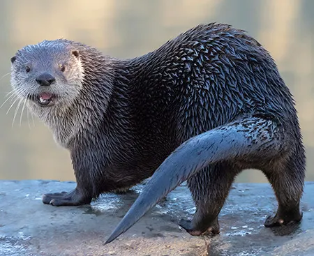
Raccoons are nocturnal and usually solitary, unless they congregate at man-made food sources such as picnic areas or campgrounds. They prefer brushy, thickly vegetated habitats, but adapt very well to the many artificial ponds, lakes, and wetlands found in the suburbs and housing developments. They can eat almost anything; their dexterous paws can easily open garbage cans, so they can readily take advantage of discarded food. In the warm desert climate, a raccoon may sleep away the day out in the open, draped over a tree branch.
Ringtails are strictly nocturnal animals, using their large eyes and keen sense of smell to locate prey. They are excellent climbers and leapers, using their long tails for balance as they negotiate steep canyon walls or trees with equal ease. The ringtails have semi-retractable claws and can rotate their hind feet 180 degrees, allowing them to descend cliffs face first. They den in niches in rock walls, boulder piles, or hollow trees. Ringtails are solitary, only pairing up for a few days of mating in April. The 2 to 4 kits are born in June. By fall the young can hunt for themselves and soon disperse. Though fierce little fighters, ringtails fall prey to great horned owls, bobcats, and coyotes. When frightened, they emit a musky odor from anal scent glands.
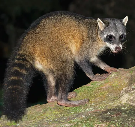
Coatis are diurnal, active mostly in the morning and late afternoon, then spending the night in trees or caves. As coatis forage through an area they travel with their long tails held vertically.
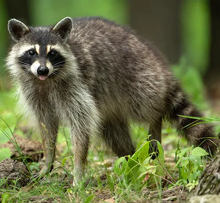
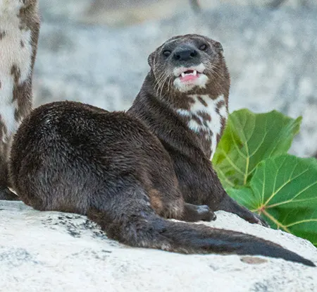
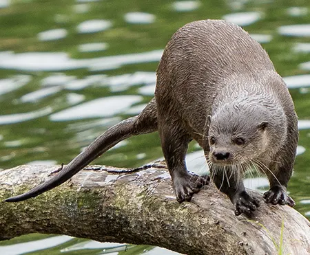
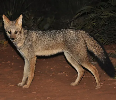
(Procyon lotor)
Raccoon
Ракун звичайний
It is found across southern Canada, throughout most of the United States, extending into northern South America, and has been introduced to parts of Asia and Europe. It lives in a variety of habitats near rivers, lakes, in floodplains, and less often in foothill deciduous and mixed forests.

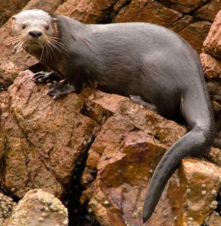
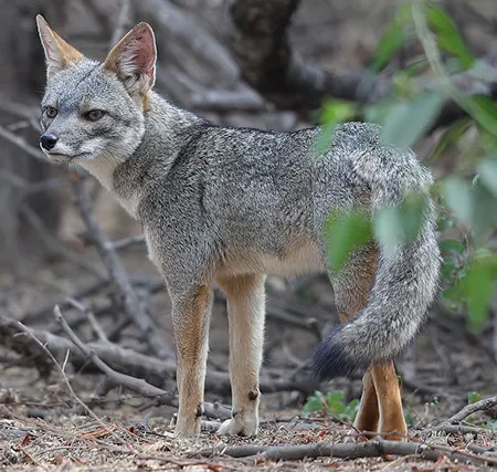
(Procyon pygmaeus)
Cozumel Raccoon
Ракун пігмейський
It is endemic to Cozumel Island, off the coast of the Yucatán Peninsula, Mexico. It inhabits a range of habitats but is primarily limited to the mangrove forests and sandy wetlands at the north-western tip of the island.
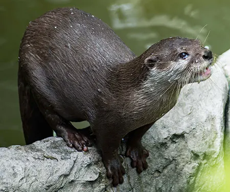
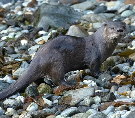
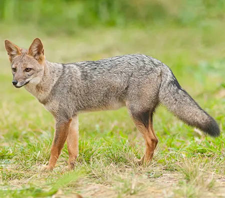
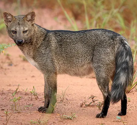
(Procyon cancrivorus)
Crab-eating Raccoon
Ракун ракоїд
It is native to marshy and jungle areas of Central and South America (including Trinidad and Tobago). It is found from Costa Rica south through most areas of South America east of the Andes down to northern Argentina and Uruguay.
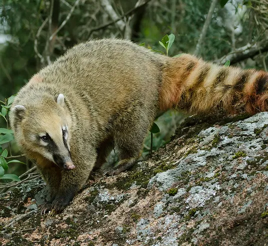
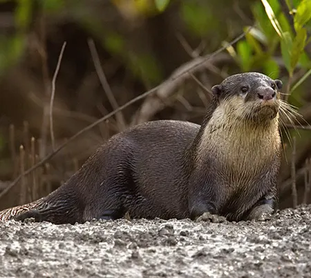
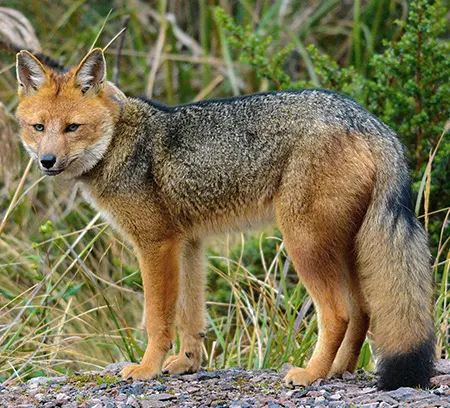
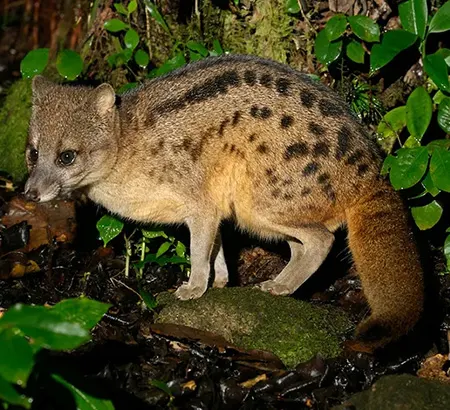
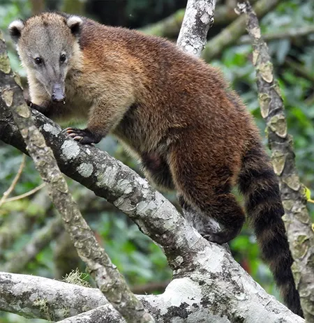
(Nasua nasua)
South American Coati
Носуха амазонська
It is widespread in tropical and subtropical South America, occurring in lowland forests east of the Andes at elevations up to 2,500 m, from Colombia and The Guianas south to Uruguay and northern Argentina.
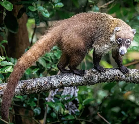
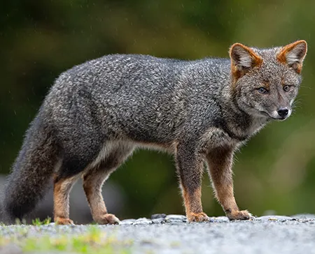
(Nasua narica)
White-nosed Coati
Носуха білоноса
It is distributed from as far north as Arizona and New Mexico, through Mexico and Central America, to the far north-western region of Colombia. It inhabits wooded areas in tropical and subtropical dry broadleaf forests and in tropical and subtropical moist broadleaf forests, at elevations from sea level to 3,000 m.
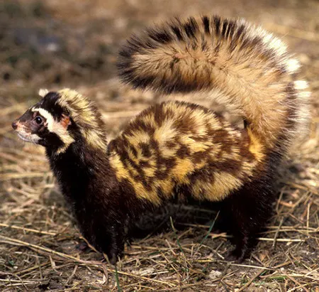
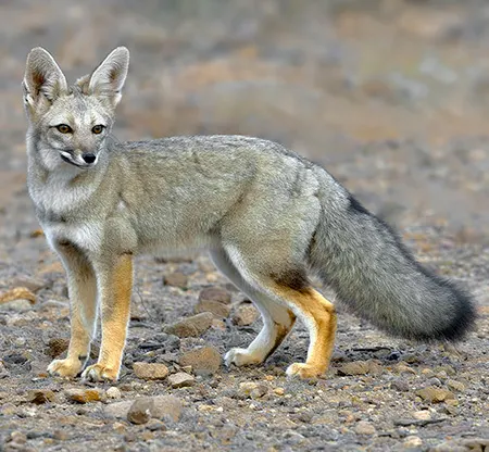
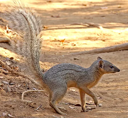
(Nasuella olivacea)
Western Mountain Coati
Західна гірська носуха
It is found in cloud forest and páramo at altitudes of 1,300–4,250 m in the Andes of Colombia and Ecuador.
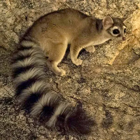
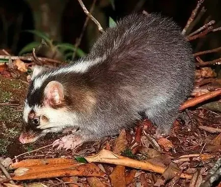
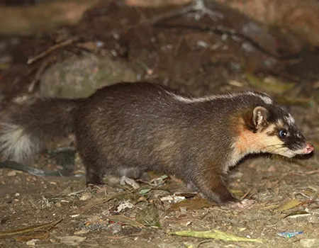
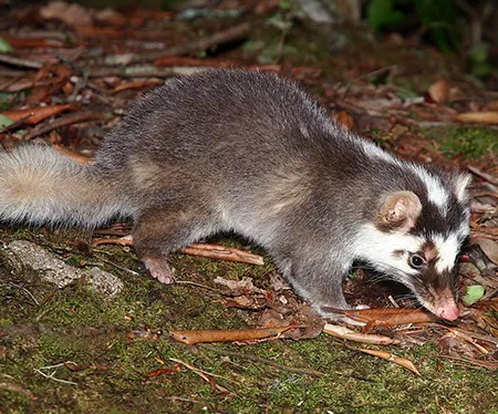
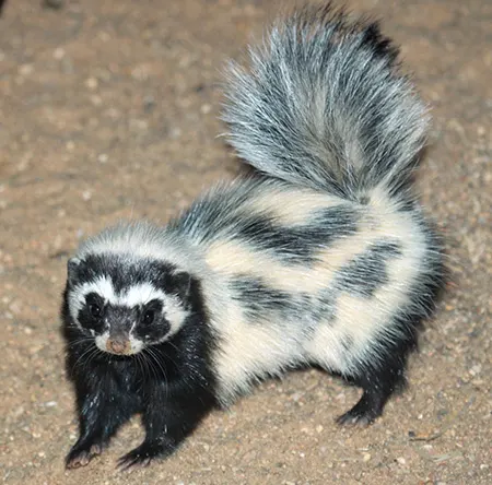
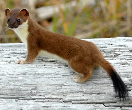
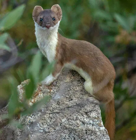
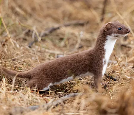
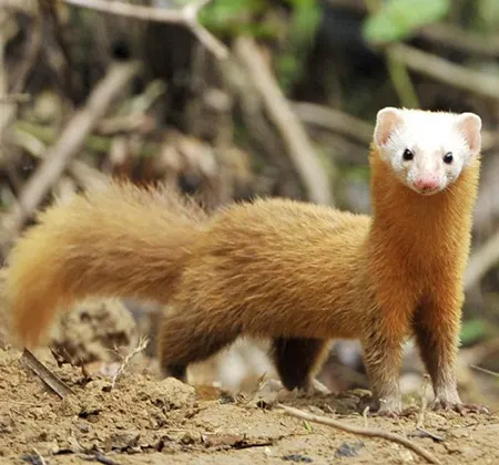
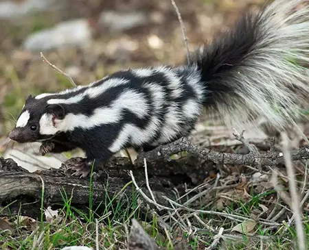
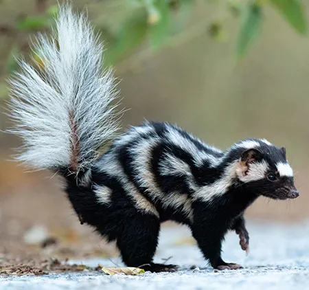
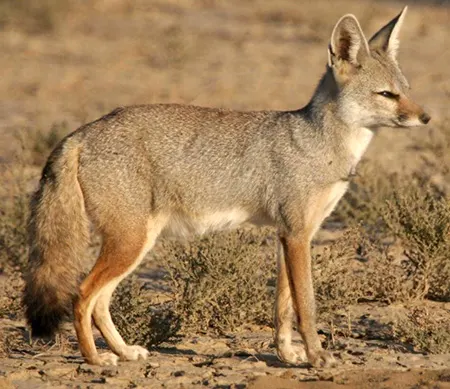
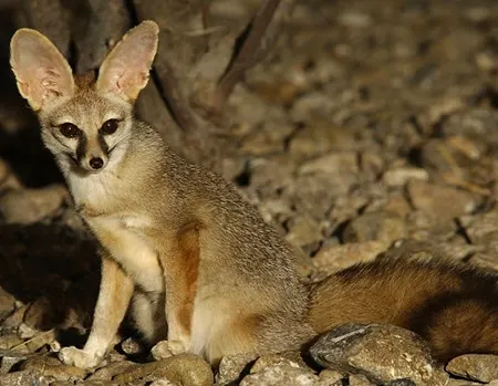
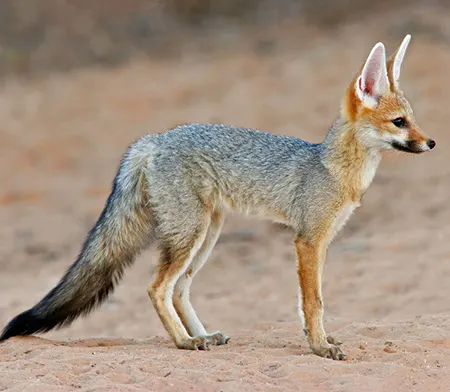
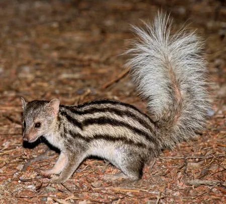
(Bassariscus astutus)
Ringtail
Котофредка смугастий
It is found in rocky desert habitats of the south-western United States and Mexico, ranging from the northern desert state of Baja California to Oaxaca.
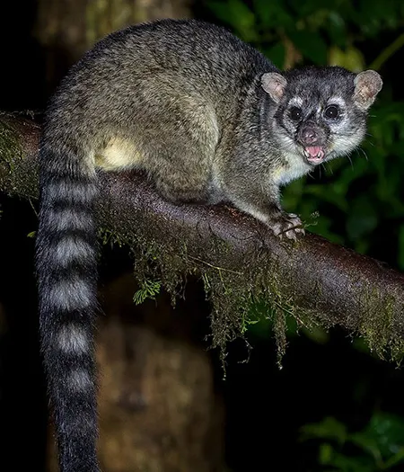
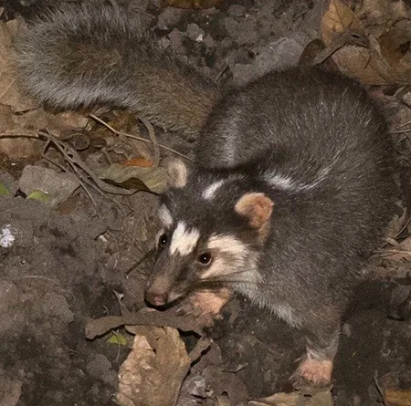
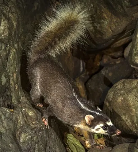
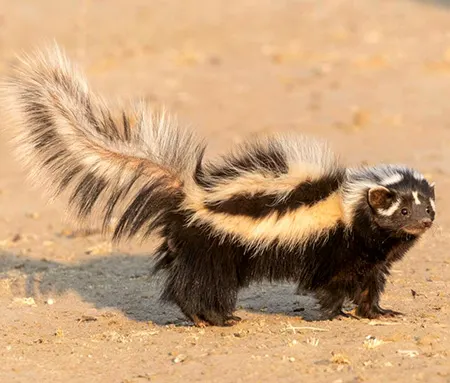
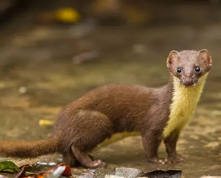
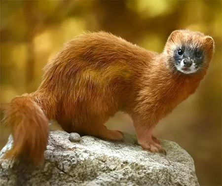
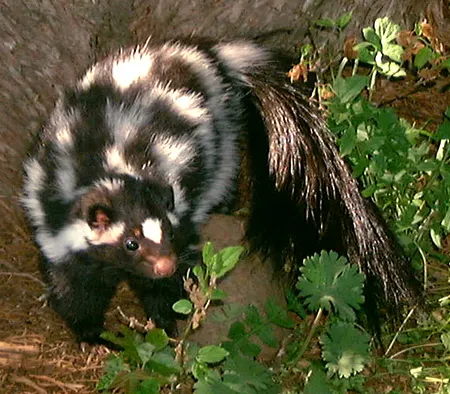
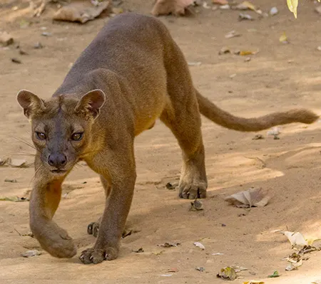
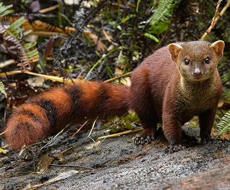
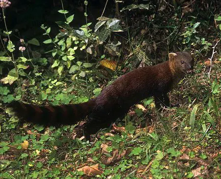
(Bassariscus sumichrasti)
Cacomistle
Какоміцл
It is fond from southern Mexico to western Panama. Its preferred habitats are humid and tropical evergreen jungle and montane cloud forests; seasonally, it may venture into drier, deciduous forests.

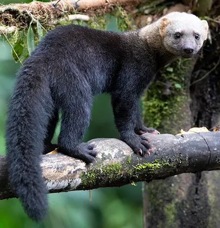
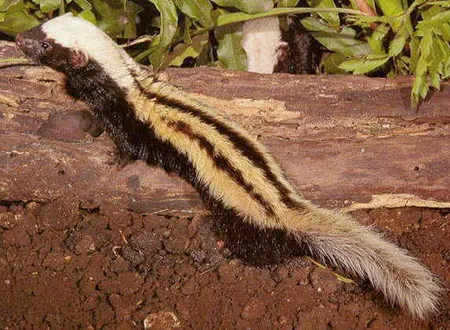
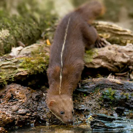
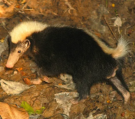
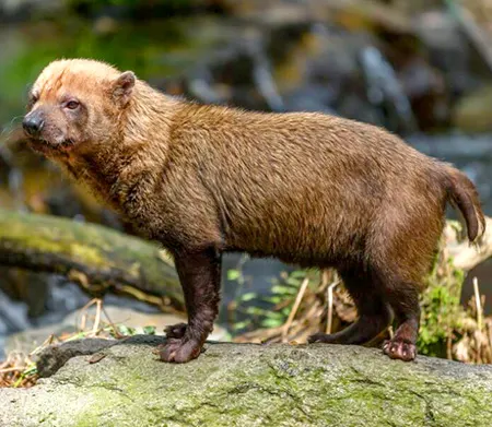
(Potos flavus)
Kinkajou
Кінкажу
It is found from southern Mexico, throughout Central America, to Bolivia east of the Andes and the Atlantic Forest of south-eastern Brazil. It is found in closed-canopy tropical forests, including lowland rainforest, montane forest, dry forest, gallery forest, and secondary forest, at elevations of up to 2,500 m.
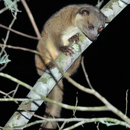
(Bassaricyon gabbii)
Northern Olingo
Олінго Ґабба
It is found from Nicaragua south through Costa Rica and Panama to Colombia. It typically inhabits montane and tropical moist forests at elevations of up to 2,000 m
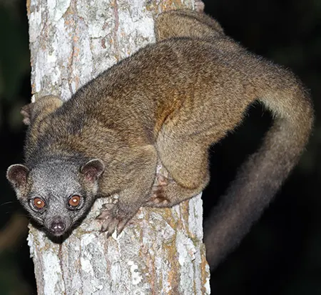
(Bassaricyon alleni)
Eastern Lowland Olingo
Олінго Алена
It it is known from the lowlands east of the Andes in Bolivia, Brazil, Colombia, Ecuador, Guyana, Peru and Venezuela.
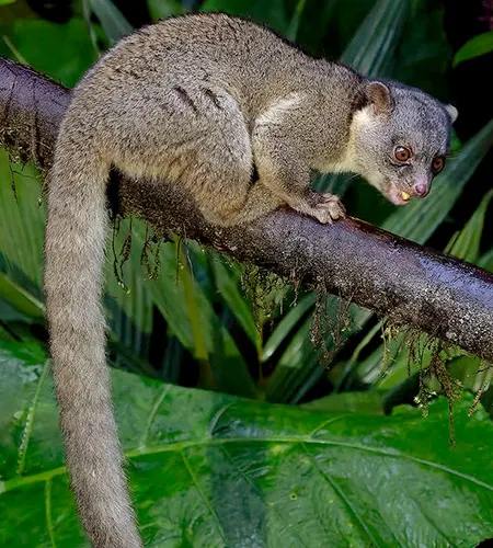
(Bassaricyon neblina)
Olinguito
Олінгіто
It is found in Andean cloud forests, ranging from western Colombia to Ecuador, at elevations between 1,500–3,000 m.
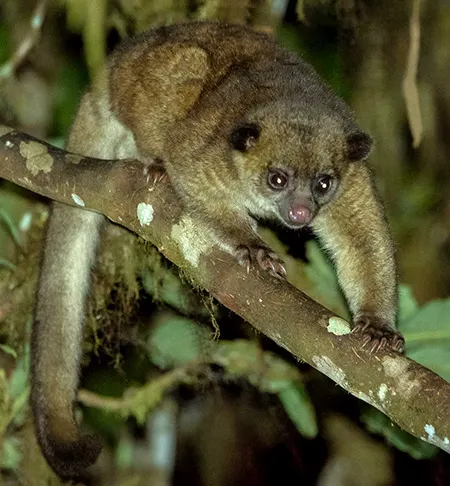
(Bassaricyon medius)
Western Lowland Olingo
Олінго західний низинний
It is found in Central and South America, specifically in Panama, Colombia and Ecuador west of the Andes.