(Reptilia)
Reptiles
Плазуни
Reptiles, as commonly defined, are a group of tetrapods with an ectothermic (‘cold-blooded’) metabolism and amniotic development. Living traditional reptiles comprise four orders: Testudines (turtles), Crocodilia (crocodilians), Squamata (lizards and snakes), and Rhynchocephalia (the tuatara). As of May 2023, about 12,000 living species of reptiles are listed in the Reptile Database.
Reptiles are tetrapod vertebrates, creatures that either have four limbs or, like snakes, are descended from four-limbed ancestors. Unlike amphibians, reptiles do not have an aquatic larval stage. Most reptiles are oviparous, although several species of squamates are viviparous, as were some extinct aquatic clades – the fetus develops within the mother, using a (non-mammalian) placenta rather than contained in an eggshell. As amniotes, reptile eggs are surrounded by membranes for protection and transport, which adapt them to reproduction on dry land. Many of the viviparous species feed their fetuses through various forms of placenta analogous to those of mammals, with some providing initial care for their hatchlings. Extant reptiles range in size from a tiny gecko, Sphaerodactylus ariasae, which can grow up to 17 mm to the saltwater crocodile, Crocodylus porosus, which can reach over 6 m in length and weigh over 1,000 kg.
Circulation
All lepidosaurs and turtles have a three-chambered heart consisting of two atria, one variably partitioned ventricle, and two aortas that lead to the systemic circulation. The degree of mixing of oxygenated and deoxygenated blood in the three-chambered heart varies depending on the species and physiological state. Under different conditions, deoxygenated blood can be shunted back to the body or oxygenated blood can be shunted back to the lungs. This variation in blood flow has been hypothesized to allow more effective thermoregulation and longer diving times for aquatic species, but has not been shown to be a fitness advantage.
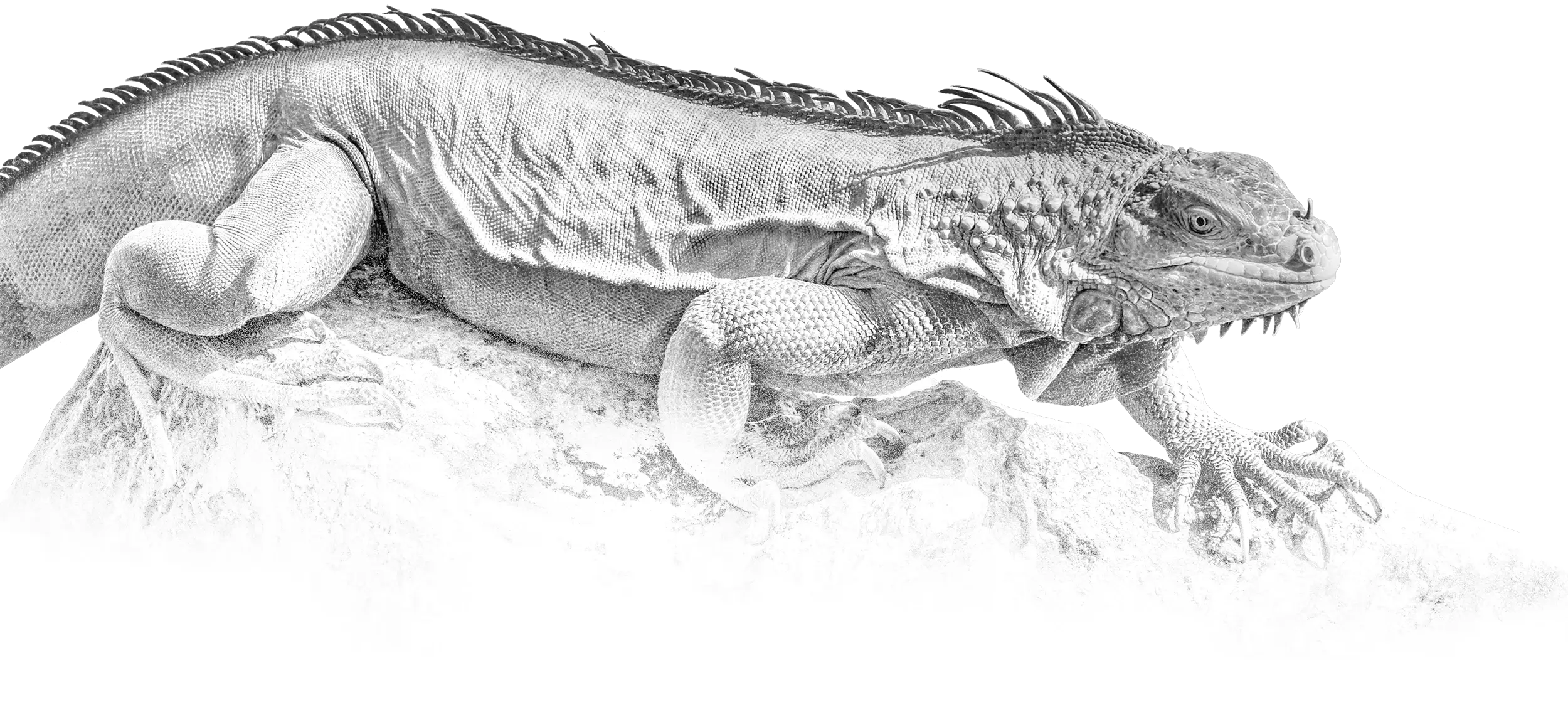
For example, iguana hearts, like the majority of the squamate hearts, are composed of three chambers with two aorta and one ventricle, cardiac involuntary muscles. The main structures of the heart are the sinus venosus, the pacemaker, the left atrium, the right atrium, the atrioventricular valve, the cavum venosum, cavum arteriosum, the cavum pulmonale, the muscular ridge, the ventricular ridge, pulmonary veins, and paired aortic arches.
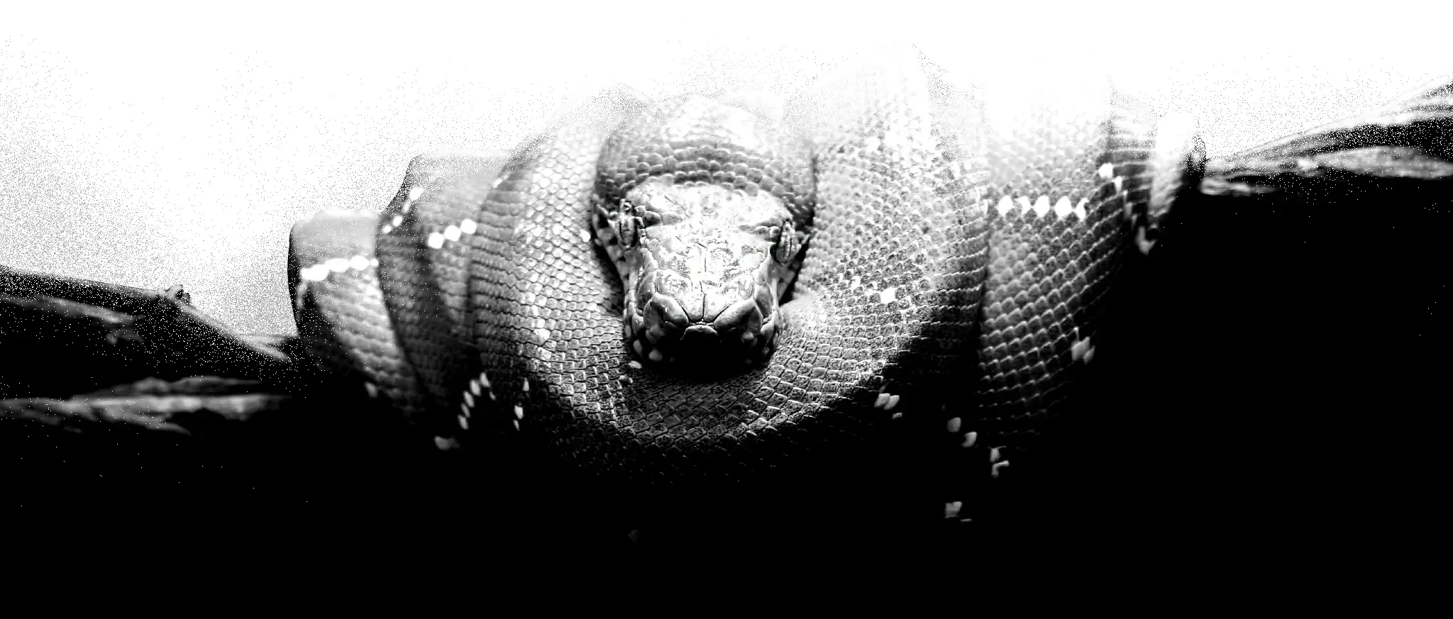
Some squamate species (e.g., pythons and monitor lizards) have three-chambered hearts that become functionally four-chambered hearts during contraction. This is made possible by a muscular ridge that subdivides the ventricle during ventricular diastole and completely divides it during ventricular systole. Because of this ridge, some of these squamates are capable of producing ventricular pressure differentials that are equivalent to those seen in mammalian and avian hearts.
Crocodilians have an anatomically four-chambered heart, similar to birds, but also have two systemic aortas and are therefore capable of bypassing their pulmonary circulation. In turtles, the ventricle is not perfectly divided, so a mix of aerated and nonaerated blood can occur.
Respiratory system
All reptiles breathe using lungs. Aquatic turtles have developed more permeable skin, and some species have modified their cloaca to increase the area for gas exchange. Even with these adaptations, breathing is never fully accomplished without lungs. Lung ventilation is accomplished differently in each main reptile group. In squamates, the lungs are ventilated almost exclusively by the axial musculature. This is also the same musculature that is used during locomotion. Because of this constraint, most squamates are forced to hold their breath during intense runs. Some, however, have found a way around it. Varanids, and a few other lizard species, employ buccal pumping as a complement to their normal “axial breathing”. This allows the animals to completely fill their lungs during intense locomotion, and thus remain aerobically active for a long time. Tegu lizards are known to possess a proto-diaphragm, which separates the pulmonary cavity from the visceral cavity. While not actually capable of movement, it does allow for greater lung inflation, by taking the weight of the viscera off the lungs.
Crocodilians actually have a muscular diaphragm that is analogous to the mammalian diaphragm. The difference is that the muscles for the crocodilian diaphragm pull the pubis (part of the pelvis, which is movable in crocodilians) back, which brings the liver down, thus freeing space for the lungs to expand. This type of diaphragmatic setup has been referred to as the “hepatic piston”. The airways form a number of double tubular chambers within each lung. On inhalation and exhalation air moves through the airways in the same direction, thus creating a unidirectional airflow through the lungs. A similar system is found in birds, monitor lizards and iguanas.
Most reptiles lack a secondary palate, meaning that they must hold their breath while swallowing. Crocodilians have evolved a bony secondary palate that allows them to continue breathing while remaining submerged (and protect their brains against damage by struggling prey). Skinks (family Scincidae) also have evolved a bony secondary palate, to varying degrees. Snakes took a different approach and extended their trachea instead. Their tracheal extension sticks out like a fleshy straw, and allows these animals to swallow large prey without suffering from asphyxiation.
How turtles breathe has been the subject of much study. To date, only a few species have been studied thoroughly enough to get an idea of how those turtles breathe. The varied results indicate that turtles have found a variety of solutions to this problem.
The difficulty is that most turtle shells are rigid and do not allow for the type of expansion and contraction that other amniotes use to ventilate their lungs. Some turtles, such as the Indian flapshell (Lissemys punctata), have a sheet of muscle that envelops the lungs. When it contracts, the turtle can exhale. When at rest, the turtle can retract the limbs into the body cavity and force air out of the lungs. When the turtle protracts its limbs, the pressure inside the lungs is reduced, and the turtle can suck air in. Turtle lungs are attached to the inside of the top of the shell (carapace), with the bottom of the lungs attached (via connective tissue) to the rest of the viscera. By using a series of special muscles (roughly equivalent to a diaphragm), turtles are capable of pushing their viscera up and down, resulting in effective respiration, since many of these muscles have attachment points in conjunction with their forelimbs (indeed, many of the muscles expand into the limb pockets during contraction).
Breathing during locomotion has been studied in three species, and they show different patterns. Adult female green sea turtles do not breathe as they crutch along their nesting beaches. They hold their breath during terrestrial locomotion and breathe in bouts as they rest. North American box turtles breathe continuously during locomotion, and the ventilation cycle is not coordinated with the limb movements. This is because they use their abdominal muscles to breathe during locomotion. The last species to have been studied is the red-eared slider, which also breathes during locomotion, but takes smaller breaths during locomotion than during small pauses between locomotor bouts, indicating that there may be mechanical interference between the limb movements and the breathing apparatus. Box turtles have also been observed to breathe while completely sealed up inside their shells.
Sound production
Compared with frogs, birds, and mammals, reptiles are less vocal. Sound production is usually limited to hissing, which is produced merely by forcing air though a partly closed glottis and is not considered to be a true vocalization. The ability to vocalize exists in crocodilians, some lizards and turtles; and typically involves vibrating fold-like structures in the larynx or glottis. Some geckos and turtles possess true vocal cords, which have elastin-rich connective tissue.
Skin
Reptilian skin is covered in a horny epidermis, making it watertight and enabling reptiles to live on dry land, in contrast to amphibians. Compared to mammalian skin, that of reptiles is rather thin and lacks the thick dermal layer that produces leather in mammals. Exposed parts of reptiles are protected by scales or scutes, sometimes with a bony base (osteoderms), forming armor. In lepidosaurs, such as lizards and snakes, the whole skin is covered in overlapping epidermal scales. Such scales were once thought to be typical of the class Reptilia as a whole, but are now known to occur only in lepidosaurs. The scales found in turtles and crocodiles are of dermal, rather than epidermal, origin and are properly termed scutes. In turtles, the body is hidden inside a hard shell composed of fused scutes.
Reptiles shed their skin through a process called ecdysis which occurs continuously throughout their lifetime. In particular, younger reptiles tend to shed once every five to six weeks while adults shed three to four times a year. Younger reptiles shed more because of their rapid growth rate. Once full size, the frequency of shedding drastically decreases. The process of ecdysis involves forming a new layer of skin under the old one. Proteolytic enzymes and lymphatic fluid is secreted between the old and new layers of skin. Consequently, this lifts the old skin from the new one allowing shedding to occur. Snakes will shed from the head to the tail while lizards shed in a “patchy pattern”. Dysecdysis, a common skin disease in snakes and lizards, will occur when ecdysis, or shedding, fails. There are numerous reasons why shedding fails and can be related to inadequate humidity and temperature, nutritional deficiencies, dehydration and traumatic injuries. Nutritional deficiencies decrease proteolytic enzymes while dehydration reduces lymphatic fluids to separate the skin layers. Traumatic injuries on the other hand, form scars that will not allow new scales to form and disrupt the process of ecdysis.
Digestion
Most reptiles are insectivorous or carnivorous and have simple and comparatively short digestive tracts due to meat being fairly simple to break down and digest. Digestion is slower than in mammals, reflecting their lower resting metabolism and their inability to divide and masticate their food. Their poikilotherm metabolism has very low energy requirements, allowing large reptiles like crocodiles and large constrictors to live from a single large meal for months, digesting it slowly.
Today, turtles are the only predominantly herbivorous reptile group, but several lines of agamas and iguanas have evolved to live wholly or partly on plants.
Herbivorous reptiles face the same problems of mastication as herbivorous mammals but, lacking the complex teeth of mammals, many species swallow rocks and pebbles (so called gastroliths) to aid in digestion: The rocks are washed around in the stomach, helping to grind up plant matter. Salt water crocodiles use gastroliths as ballast, stabilizing them in the water or helping them to dive. A dual function as both stabilizing ballast and digestion aid has been suggested for gastroliths found in plesiosaurs.
Nerves
The reptilian nervous system contains the same basic part of the amphibian brain, but the reptile cerebrum and cerebellum are slightly larger. Most typical sense organs are well developed with certain exceptions, most notably the snake’s lack of external ears (middle and inner ears are present). There are twelve pairs of cranial nerves. Due to their short cochlea, reptiles use electrical tuning to expand their range of audible frequencies.
Vision
Most reptiles are diurnal animals. The vision is typically adapted to daylight conditions, with color vision and more advanced visual depth perception than in amphibians and most mammals.
Reptiles usually have excellent vision, allowing them to detect shapes and motions at long distances. They often have poor vision in low-light conditions. Birds, crocodiles and turtles have three types of photoreceptor: rods, single cones and double cones, which gives them sharp color vision and enables them to see ultraviolet wavelengths. The lepidosaurs appear to have lost the duplex retina and only have a single class of receptor that is cone-like or rod-like depending on whether the species is diurnal or nocturnal. In many burrowing species, such as blind snakes, vision is reduced.
Many lepidosaurs have a photosensory organ on the top of their heads called the parietal eye, which are also called third eye, pineal eye or pineal gland. This “eye” does not work the same way as a normal eye does as it has only a rudimentary retina and lens and thus, cannot form images. It is, however, sensitive to changes in light and dark and can detect movement.
Some snakes have extra sets of visual organs (in the loosest sense of the word) in the form of pits sensitive to infrared radiation (heat). Such heat-sensitive pits are particularly well developed in the pit vipers, but are also found in boas and pythons. These pits allow the snakes to sense the body heat of birds and mammals, enabling pit vipers to hunt rodents in the dark.
Most reptiles, as well as birds, possess a nictitating membrane, a translucent third eyelid which is drawn over the eye from the inner corner. In crocodilians, it protects its eyeball surface while allowing a degree of vision underwater. However, many squamates, geckos and snakes in particular, lack eyelids, which are replaced by a transparent scale. This is called the brille, spectacle, or eyecap. The brille is usually not visible, except for when the snake molts, and it protects the eyes from dust and dirt.
Reproduction
Reptiles generally reproduce sexually, though some are capable of asexual reproduction. All reproductive activity occurs through the cloaca, the single exit/entrance at the base of the tail where waste is also eliminated. Most reptiles have copulatory organs, which are usually retracted or inverted and stored inside the body. In turtles and crocodilians, the male has a single median penis, while squamates, including snakes and lizards, possess a pair of hemipenes, only one of which is typically used in each session. Tuatara, however, lack copulatory organs, and so the male and female simply press their cloacas together as the male discharges sperm.
Most reptiles lay amniotic eggs covered with leathery or calcareous shells. An amnion, chorion, and allantois are present during embryonic life. The eggshell protects the crocodile embryo and keeps it from drying out, but it is flexible to allow gas exchange. The chorion aids in gas exchange between the inside and outside of the egg. It allows carbon dioxide to exit the egg and oxygen gas to enter the egg. The albumin further protects the embryo and serves as a reservoir for water and protein. The allantois is a sac that collects the metabolic waste produced by the embryo. The amniotic sac contains amniotic fluid which protects and cushions the embryo. The amnion aids in osmoregulation and serves as a saltwater reservoir. The yolk sac surrounding the yolk contains protein and fat rich nutrients that are absorbed by the embryo via vessels that allow the embryo to grow and metabolize. The air space provides the embryo with oxygen while it is hatching. This ensures that the embryo will not suffocate while it is hatching. There are no larval stages of development. Viviparity and ovoviviparity have evolved in squamates and many extinct clades of reptiles. Among squamates, many species, including all boas and most vipers, use this mode of reproduction. The degree of viviparity varies; some species simply retain the eggs until just before hatching, others provide maternal nourishment to supplement the yolk, and yet others lack any yolk and provide all nutrients via a structure similar to the mammalian placenta.
Asexual reproduction has been identified in squamates in six families of lizards and one snake. In some species of squamates, a population of females is able to produce a unisexual diploid clone of the mother. This form of asexual reproduction, called parthenogenesis, occurs in several species of gecko, and is particularly widespread in the teiids (especially Aspidocelis) and lacertids (Lacerta). In captivity, Komodo dragons (Varanidae) have reproduced by parthenogenesis.
Parthenogenetic species are suspected to occur among chameleons, agamids, xantusiids, and typhlopids.
Some reptiles exhibit temperature-dependent sex determination, in which the incubation temperature determines whether a particular egg hatches as male or female. This type of sex determination is most common in turtles and crocodiles, but also occurs in lizards and tuatara.
Defense mechanisms
Many small reptiles, such as snakes and lizards, that live on the ground or in the water are vulnerable to being preyed on by all kinds of carnivorous animals. Thus, avoidance is the most common form of defense in reptiles. At the first sign of danger, most snakes and lizards crawl away into the undergrowth, and turtles and crocodiles will plunge into water and sink out of sight.
Reptiles tend to avoid confrontation through camouflage. Two major groups of reptile predators are birds and other reptiles, both of which have well-developed color vision. Thus the skins of many reptiles have cryptic coloration of plain or mottled gray, green, and brown to allow them to blend into the background of their natural environment. Aided by the reptiles’ capacity for remaining motionless for long periods, the camouflage of many snakes is so effective that people or domestic animals are most typically bitten because they accidentally step on them.
When camouflage fails to protect them, blue-tongued skinks will try to ward off attackers by displaying their blue tongues, and the frill-necked lizard will display its brightly colored frill. These same displays are used in territorial disputes and during courtship. If danger arises so suddenly that flight is useless, crocodiles, turtles, some lizards, and some snakes hiss loudly when confronted by an enemy. Rattlesnakes rapidly vibrate the tip of the tail, which is composed of a series of nested, hollow beads to ward off approaching danger.
In contrast to the normal drab coloration of most reptiles, the lizards of the genus Heloderma (the Gila monster and the beaded lizard) and many of the coral snakes have high-contrast warning coloration, warning potential predators they are venomous. A number of non-venomous North American snake species have colorful markings similar to those of the coral snake, an oft cited example of Batesian mimicry.
Camouflage does not always fool a predator. When caught out, snake species adopt different defensive tactics and use a complicated set of behaviors when attacked. Some species, like cobras or hognose snakes, first elevate their head and spread out the skin of their neck in an effort to look large and threatening. Failure of this strategy may lead to other measures practiced particularly by cobras, vipers, and closely related species, which use venom to attack. The venom is modified saliva, delivered through fangs from a venom gland. Some non-venomous snakes, such as American hognose snakes or European grass snake, play dead when in danger; some, including the grass snake, exude a foul-smelling liquid to deter attackers.
When a crocodilian is concerned about its safety, it will gape to expose the teeth and tongue. If this does not work, the crocodilian gets a little more agitated and typically begins to make hissing sounds. After this, the crocodilian will start to change its posture dramatically to make itself look more intimidating. The body is inflated to increase apparent size. If absolutely necessary, it may decide to attack an enemy.
Geckos, skinks, and some other lizards that are captured by the tail will shed part of the tail structure through a process called autotomy and thus be able to flee. The detached tail will continue to thrash, creating a deceptive sense of continued struggle and distracting the predator’s attention from the fleeing prey animal. The detached tails of leopard geckos can wiggle for up to 20 minutes. The tail grows back in most species, but some, like crested geckos, lose their tails for the rest of their lives. In many species the tails are of a separate and dramatically more intense color than the rest of the body so as to encourage potential predators to strike for the tail first. In the shingleback skink and some species of geckos, the tail is short and broad and resembles the head, so that the predators may attack it rather than the more vulnerable front part.
Reptiles that are capable of shedding their tails can partially regenerate them over a period of weeks. The new section will however contain cartilage rather than bone, and will never grow to the same length as the original tail. It is often also distinctly discolored compared to the rest of the body and may lack some of the external sculpting features seen in the original tail.
Source: Wikipedia

